Key to Genera in the Subfamilies Diamesinae, Prodiamesinae, Podonominae and Orthocladiinae
Ian R. Walker

Recommended Citation:
Walker, I. R.,
1997. Key to genera in the subfamilies Diamesinae, Prodiamesinae, Podonominae and Orthocladiinae Part III. In: I. R.
Walker (Ed.), The WWW Field Guide to Subfossil Midges. (http://www.sci.ouc.bc.ca/fwsc/iwalker/chironomid/orthocladiinae/orthkey3.htm)
Last Update: 11 June 1997
©1997 Ian R. Walker. ALL RIGHTS RESERVED
- a) Ventromental plates completely overlapping all pairs of lateral teeth (Fig.
1)
.............................................................................Potthastia?
- b) Ventromental plates smaller, some lateral mental teeth may extend beyond anterior ventromental margin of flattened head capsule (Fig.
2)
.............................................................................2.
- a) Ventromental plates wholly or partially overlapping some mental teeth, with approximately straight or weakly concave antero-lateral margins; median tooth dark; premandible simple (Fig. 2)
.............................................................................Psectrocladius (Monopsectrocladius) type
- b) Ventromental plates not overlapping mental teeth (Fig.
4); or if wholly or partially overlapping mental teeth, then anterolateral margin convex, or median tooth with little or no pigmentation; premandible simple or compound (Fig.
8)
.............................................................................3.
- a) Ventromental plates very broad in submental region
(Fig. 3).
.............................................................................Stilocladius
- b) Ventromental plates not exceptionally broad in submental region (Fig. 6)
.............................................................................4.
- a) Median tooth flanked by 2 pairs of light-coloured lateral teeth (Fig.
4)
............................................................................."Cricotopus" type
- b) Median tooth flanked by 1 pair of light-colured lateral teeth, or all lateral teeth similarly pigmented (Fig.
5)
.............................................................................5.
- a) Median tooth weakly notched laterally (Fig. 5)
.............................................................................Parakiefferiella sp. B
- b) Median tooth unnotched (Fig.
6)
.............................................................................6.
- a) Second lateral teeth distinctly smaller than adjacent teeth (but 1st and 2nd lateral teeth absent when mouthparts strongly worn); median tooth as dark as lateral teeth (Fig. 6)
.............................................................................Parakiefferiella nigra
- b) Second lateral teeth of similar size to adjacent pairs; median tooth with light or dark pigmentation (Fig.
9)
.............................................................................7.
- a) Median tooth very broad and with little, if any, pigmentation
(Fig. 9).
.............................................................................8.
- b) Median tooth less broad, with distinct pigmentation (varying from pale yellow or tan to dark brown) (Fig. 7)
.............................................................................Smittia/Pseudosmittia type
- a) Median tooth strongly arched
(Fig. 8).
.............................................................................Parakiefferiella triquetra type
- b) Median tooth weakly arched (Fig. 9)
.............................................................................Paracladius type
Fig. 1. Potthastia?.

Fig. 2. Psectrocladius (Monopsectrocladius)
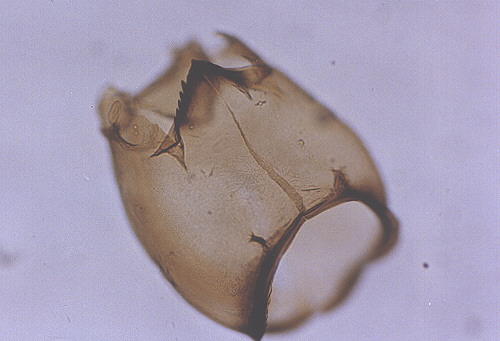
Fig. 3. Stilocladius.

Fig. 4. "Cricotopus" type.
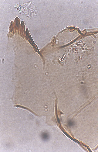
Fig. 5. Parakiefferiella sp. B.
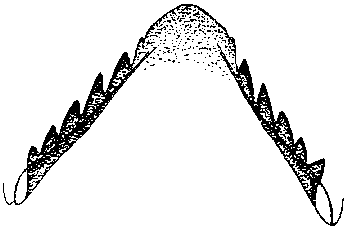
Fig. 6. Parakiefferiella nigra.
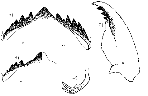
Fig. 7. Smittia/Pseudosmittia type.

Fig. 8. Parakiefferiella triquetra type.
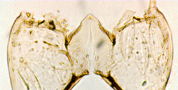
Fig. 9. Paracladius.
